All About Coffee by William H. Ukers (interesting novels in english TXT) 📖

- Author: William H. Ukers
- Performer: -
Book online «All About Coffee by William H. Ukers (interesting novels in english TXT) 📖». Author William H. Ukers
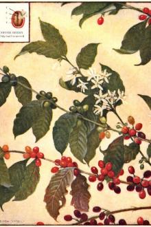
The coffee bean with which the consumer is familiar is only a small part of the fruit. The fruit, which is the size of a small cherry, has, like the cherry, an outer fleshy portion called the pericarp. Beneath this is a part like tissue paper, spoken of technically as the parchment, but known scientifically as the endocarp. Next in position to this, and covering the seed, is the so-called spermoderm, which means the seed skin, referred to in the trade as the silver skin. Small portions of this silver skin are always to be found in the cleft of the coffee bean.
The coffee bean is the embryo and its food supply; the embryo is that part of the seed which, when supplied with food and moisture, develops into a new plant. The embryo of the coffee is very minute (Fig. 331, II, Em)[101]; and the greater part of the seed is taken up by the food supply, consisting of hard and soft endosperm (Fig. 331, I and II, Sa, Sp). The minute embryo consists of two small thick leaves, the cotyledons (Fig. 331, III, cot), a short stem, invisible in the undissected embryo, and a small root, the radicle (Fig. 331, III, rad).
Fig. 332. Coffee. Cross section of beanshowing folded endosperm with hard and soft tissues. x6. (Moeller)
Fruit Structure
In order to examine the structure of these layers of the fruit under the microscope, it is necessary to use the pericarp dry, as it is not easily obtainable in its natural condition. If desired, an alcoholic specimen may be used, but it has been found that the dry method gives more satisfactory results. The dried pericarp is about 0.5 mm thick. Great difficulty is experienced in cutting microtome sections of pericarp when the specimen is embedded in paraffin, because the outer layers are soft and the endocarp is hard, and the two parts of the section separate at this point. To overcome this, the sections might also be embedded in celloidin. When the sections are satisfactory, they may be stained with any of the double stains ordinarily used in the study of plant histology.
Fig. 333. Coffee. Cross section of hull and bean. Pericarp consists of: 1, epicarp; 2–3, layers of mesocarp, with 4, fibro-vascular bundle; 5, palisade layer; and 6, endocarp; ss, spermoderm, consists of 8, sclerenchyma, and 9, parenchyma; End, endosperm (Tschirch and Oesterle)
A section cut crosswise through the entire fruit would present the appearance shown in Fig. 333. The cells of the epicarp are broad and polygonal, sometimes regularly four-sided, about 15–35 µ broad. At intervals along the surface of the epicarp are stomata, or breathing pores, surrounded by guard cells. The next layer of the pericarp is the mesocarp (Figs. 333, 334, 335), the cells of which are larger and more regular in outline than the epicarp. The cells of the mesocarp become as large as 100 µ broad, but in the inner parts of the layer they become very much flattened. Fibrovascular bundles are scattered through the compressed cells of the mesocarp. The cell walls are thick; and large, amorphous, brown masses are found within the cell; occasionally, large crystals are found in the outer part of the layer. The fibro-vascular bundles consist mainly of bast and wood fibers and vessels. The bast fibers are as large as 1 mm long and 25 µ broad, with thick walls and very small lumina. Spiral and pitted vessels are also present.
Fig. 334. Coffee. Surface view of ep, epicarp, and p, outer parenchyma of mesocarp. x160. (Moeller)
The layer next to this is a soft tissue, parenchyma (Fig. 333, 5; Fig. 334, p). The parenchyma, or palisade cells as they are called, is a thin-walled tissue in which the cells are elongated, from which fact they receive their name. The walls of these cells, though very thin, are mucilaginous, and capable of taking up large amounts of water. They stain well with the aniline stains.
The endocarp (Fig. 336) is closely connected with the palisade layer and has thin-walled cells that closely resemble, in all respects, the endocarp of the apple. The outer layer consists of thick-walled fibers, which are remarkably porous (Fig. 333, 6; Fig. 336) while the fibers of the inner layer are thin-walled and run in the transverse direction.
The Bean Structure
Spermoderm, or silver skin, is not difficult to secure for microscopic analysis; because shreds of it remain in the groove of the berry, and these shreds are ample for examination. It can readily be removed without tearing, if soaked in water for a few hours. The spermoderm is thin enough not to need sectioning. It consists of two elements—sclerenchyma and parenchyma cells. (Figs. 333, 337, st, p).
Fig. 335. Coffee. Elements of pericarp in surface view. p, parenchyma; bp, parenchyma of fibro-vascular bundle; b, bast fiber; sp, spiral vessel. x160. (Moeller)
Sclerenchyma forms an uninterrupted covering in the early stages of the seed; but as the seed develops, surrounding tissues grow more rapidly than the sclerenchyma, and the cells are pushed apart and scattered. The cells occurring in the cleft of the berry are straight, narrow, and long, becoming as long as 1 mm, and resemble bast fibers somewhat. On the surface of the berry, and sometimes in the cleft, there are found smaller, thicker cells, which are irregular in outline, club-shaped and vermiform types predominating.
Parenchyma cells form the remainder of the spermoderm; and these are partially obliterated, so that the structure is not easily seen, appearing almost like a solid membrane. The raphe runs through the parenchyma found in the cleft of the berry.
The endosperm (Figs. 333; 338) consist of small cells in the outer part, and large cells, frequently as thick as 100 µ, in the inner part. The cell walls are thickened and knotted. Certain of the inner cells have mucilaginous walls which when treated with water disappear, leaving only the middle lamellae, which gives the section a peculiar appearance. The cells contain no starch, the reserve food supply being stored cellulose, protein, and aleurone grains. Various investigators report the presence of sugar, tannin, iron, salts, and caffein.
The embryo (Fig. 331, III) may be obtained by soaking the bean in water for several hours, cutting through the cleft and carefully breaking apart the endosperm. If it is now soaked in diluted alkali, the embryo protrudes through the lower end of the endosperm. It is then cleared in alkali, or in chloral hydrate. The cotyledons shown have three pairs of veins, which are slightly netted. The radicle is blunt and is about 3⁄4 mm in length, while the cotyledons are 1⁄2 mm long.
Fig. 336. Coffee. Sclerenchyma fibers of endocarp. x160. (Moeller)
The Coffee-Leaf Disease
The coffee tree has many pests and diseases; but the disease most feared by planters is that generally referred to as the coffee-leaf disease, and by this is meant the fungoid Hemileia vastatrix, which as told in chapter XV, destroyed Ceylon's once prosperous coffee industry. As it has since been found in nearly all coffee-producing countries, it has become a nightmare in the dreams of all coffee planters. The microscope shows how the spores of this dreaded fungus, carried by the winds upon a leaf of the coffee tree, proceed to germinate at the expense of the leaf; robbing it of its nourishment, and causing it to droop and to die. A mixture of powdered lime and sulphur has been found to be an effective germicide, if used in time and diligently applied.
Fig. 337. Coffee. Spermoderm in surface view. st. sclerenchyma; p, compressed parenchyma. x160. (Moeller)
Fig. 338. Coffee. Cross-section of outer layers of endosperm, showing knotty thickenings of cell walls. x160. (Moeller)
Fig. 339. Coffee. Tissues of embryo in section. x160. (Moeller)
Value of Microscopic Analysis
The value of the microscopic analysis of coffee may not be apparent at first sight; but when one realizes that in many cases the microscopic examination is the only way to detect adulteration in coffee, its importance at once becomes apparent. In many instances the chemical analysis fails to get at the root of the trouble, and then the only method to which the tester has recourse is the examination of the suspected material under the scope. The mixing of chicory with coffee has in the past been one of the commonest forms of adulteration. The microscopic examination in this connection is the most reliable. The coffee grain will have the appearance already described. Microscopically, chicory shows numerous thin-walled parenchymatous cells, lactiferous vessels, and sieve tubes with transverse plates. There are also present large vessels with huge, well-defined pits.
1. under surface of affected leaf, x 1⁄2; 2, section through same showing mycelium, haustoria, and a spore-cluster; 3, a spore-cluster seen from below; 4, a uredospore; 5, germinating uredospore; 6, appressorial swellings at tips of germ-tubes; 7, infection through stoma of leaf; 8, teleutospores; 9, teleutospore germinating with promycelium and sporidia; 10, sporidia and their germination (2 after Zimmermann, 3 after Delacroix, 4–10 after Ward)
Roasted date stones have been used as adulterants, and these can be detected quite readily with the aid of the microscope, as they have a very characteristic microscopic appearance. The epidermal cells are almost oblong, while the parenchymatous cells are large, irregular and contain large quantities of tannin.
Adulteration and adulterants are considered more fully in chapter XVII.
Green bean, showing the size and form of the cells as well as the drops of oil contained within their cavities. Drawn with the camera lucida, and magnified 140 diameters.
A fragment of roasted coffee under the microscope. Drawn with the camera lucida, and magnified 140 diameters.
Green and Roasted Coffee Under the Microscope
Longitudinal—Magnified 200 diameters
Cross Section—Magnified 200 diameters
Tangential—Magnified 200 diameters
Tangential—Magnified 200 diameters
GREEN AND ROASTED BOGOTA COFFEE UNDER THE MICROSCOPE
These pictures serve to demonstrate that the coffee bean is made up of minute cells that are not broken down to any extent by the roasting process. Note that the oil globules are more prominent in the green than in the roasted product
Chapter XVII THE CHEMISTRY OF THE COFFEE BEANChemistry of the preparation and treatment of the green bean—Artificial aging—Renovating damaged coffees—Extracts—"Caffetannic acid"—Caffein, caffein-free coffee—Caffeol—Fats and oils—Carbohydrates—Roasting—Scientific aspects of grinding and packaging—The coffee brew—Soluble coffee—Adulterants and substitutes—Official methods of analysis
By Charles W. Trigg
Industrial Fellow of the Mellon Institute of Industrial Research, Pittsburgh, 1916–1920
When the vast


Comments (0)